View in English?
View in English?
View in English?
| Cat. # | Size | Qty. | Price | Inventory |
|---|---|---|---|---|
| 2130S | 100 µl |
|
| REACTIVITY | H |
| SENSITIVITY | Endogenous |
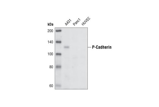
| MW (kDa) | 120 |
| SOURCE | Rabbit |
Product Information
| Application | Dilution |
|---|---|
| Western Blotting | 1:1000 |
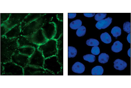
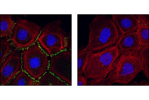
| Immunofluorescence (Immunocytochemistry) | 1:100 |
For western blots, incubate membrane with diluted primary antibody in 5% w/v BSA, 1X TBS, 0.1% Tween® 20 at 4°C with gentle shaking, overnight.
NOTE: Please refer to primary antibody product webpage for recommended antibody dilution.
From sample preparation to detection, the reagents you need for your Western Blot are now in one convenient kit: #12957 Western Blotting Application Solutions Kit
NOTE: Prepare solutions with reverse osmosis deionized (RODI) or equivalent grade water.
Load 20 µl onto SDS-PAGE gel (10 cm x 10 cm).
NOTE: Loading of prestained molecular weight markers (#59329, 10 µl/lane) to verify electrotransfer and biotinylated protein ladder (#7727, 10 µl/lane) to determine molecular weights are recommended.
NOTE: Volumes are for 10 cm x 10 cm (100 cm2) of membrane; for different sized membranes, adjust volumes accordingly.
* Avoid repeated exposure to skin.
posted June 2005
revised June 2020
Protocol Id: 10
Achieve higher quality immunofluorescent images using the efficient and cost-effective, pre-made reagents in our #12727 Immunofluorescence Application Solutions Kit
NOTE: Prepare solutions with reverse osmosis deionized (RODI) or equivalent grade water.
Recommended Fluorochrome-conjugated Anti-Rabbit secondary antibodies:
NOTE: Cells should be grown, treated, fixed and stained directly in multi-well plates, chamber slides or on coverslips.
Aspirate liquid, then cover cells to a depth of 2–3 mm with 4% formaldehyde diluted in 1X PBS.
NOTE: Formaldehyde is toxic, use only in a fume hood.
NOTE: All subsequent incubations should be carried out at room temperature unless otherwise noted in a humid light-tight box or covered dish/plate to prevent drying and fluorochrome fading.
posted November 2006
revised November 2013
Protocol Id: 24
Human
Polyclonal antibodies are produced by immunizing animals with a synthetic peptide corresponding to human P-cadherin. Antibodies are purified using protein A and peptide affinity chromatography.
Cadherins are a superfamily of transmembrane glycoproteins that contain cadherin repeats of approximately 100 residues in their extracellular domain. Cadherins mediate calcium-dependent cell-cell adhesion and play critical roles in normal tissue development (1). The classic cadherin subfamily includes N-, P-, R-, B-, and E-cadherins, as well as about ten other members that are found in adherens junctions, a cellular structure near the apical surface of polarized epithelial cells. The cytoplasmic domain of classical cadherins interacts with β-catenin, γ-catenin (also called plakoglobin), and p120 catenin. β-catenin and γ-catenin associate with α-catenin, which links the cadherin-catenin complex to the actin cytoskeleton (1,2). While β- and γ-catenin play structural roles in the junctional complex, p120 regulates cadherin adhesive activity and trafficking (1-4). Investigators consider E-cadherin an active suppressor of invasion and growth of many epithelial cancers (1-3). Research studies indicate that cancer cells have upregulated N-cadherin in addition to loss of E-cadherin. This change in cadherin expression is called the "cadherin switch." N-cadherin cooperates with the FGF receptor, leading to overexpression of MMP-9 and cellular invasion (3). Research studies have shown that in endothelial cells, VE-cadherin signaling, expression, and localization correlate with vascular permeability and tumor angiogenesis (5,6). Investigators have also demonstrated that expression of P-cadherin, which is normally present in epithelial cells, is also altered in ovarian and other human cancers (7,8).
Explore pathways related to this product.
STRING - Known and Predicted Protein-Protein Interactions.
Except as otherwise expressly agreed in a writing signed by a legally authorized representative of CST, the following terms apply to Products provided by CST, its affiliates or its distributors. Any Customer's terms and conditions that are in addition to, or different from, those contained herein, unless separately accepted in writing by a legally authorized representative of CST, are rejected and are of no force or effect.
Products are labeled with For Research Use Only or a similar labeling statement and have not been approved, cleared, or licensed by the FDA or other regulatory foreign or domestic entity, for any purpose. Customer shall not use any Product for any diagnostic or therapeutic purpose, or otherwise in any manner that conflicts with its labeling statement. Products sold or licensed by CST are provided for Customer as the end-user and solely for research and development uses. Any use of Product for diagnostic, prophylactic or therapeutic purposes, or any purchase of Product for resale (alone or as a component) or other commercial purpose, requires a separate license from CST. Customer shall (a) not sell, license, loan, donate or otherwise transfer or make available any Product to any third party, whether alone or in combination with other materials, or use the Products to manufacture any commercial products, (b) not copy, modify, reverse engineer, decompile, disassemble or otherwise attempt to discover the underlying structure or technology of the Products, or use the Products for the purpose of developing any products or services that would compete with CST products or services, (c) not alter or remove from the Products any trademarks, trade names, logos, patent or copyright notices or markings, (d) use the Products solely in accordance with CST Product Terms of Sale and any applicable documentation, and (e) comply with any license, terms of service or similar agreement with respect to any third party products or services used by Customer in connection with the Products.
View in English?