View in English?
View in English?
View in English?

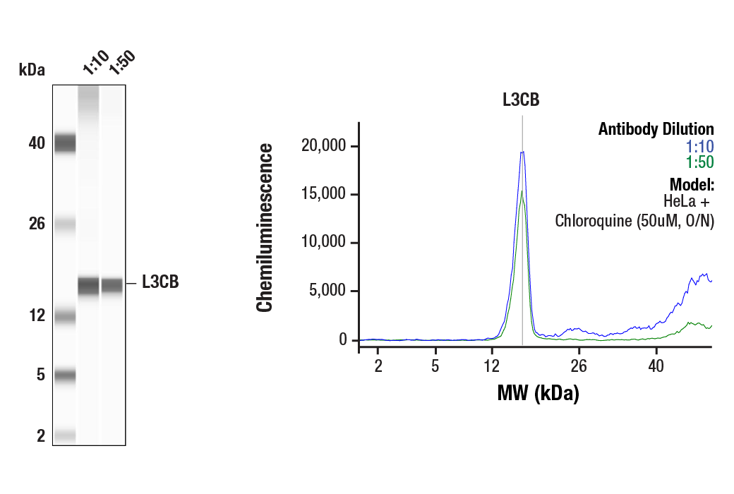
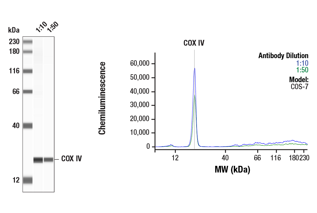
Simple Western™ analysis of lysates (1mg/ml) from HeLa cells treated with Chloroquine (50uM, O/N) using LC3B (D11) XP® Rabbit mAb #3868. The virtual lane view (left) shows the target band (as indicated) at 1:10 and 1:50 dilutions of primary antibody. The corresponding electropherogram view (right) plots chemiluminescence by molecular weight along the capillary at 1:10 (blue line) and 1:50 (green line) dilutions of primary antibody. This experiment was performed under reducing conditions on the Jess™ Simple Western instrument from ProteinSimple, a BioTechne brand, using the 2-40kDa.
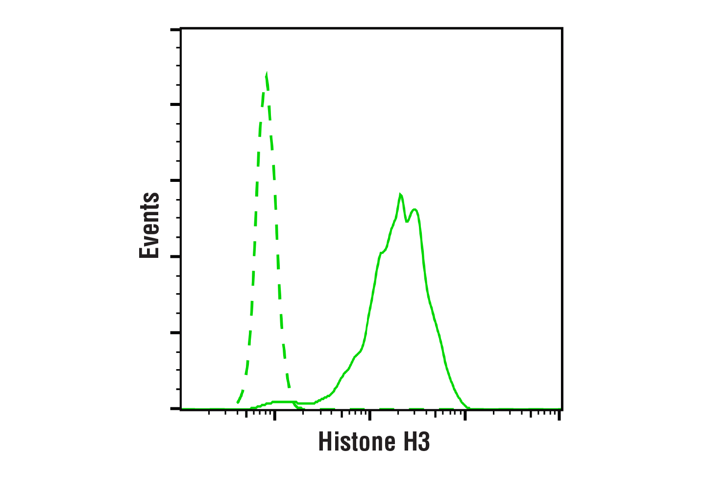
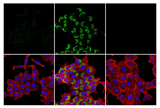
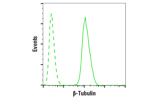
Flow cytometric analysis of K-562 cells using β-Tubulin (9F3) Rabbit mAb (solid line) compared to concentration-matched Rabbit (DA1E) mAb IgG XP® Isotype Control #3900 (dashed line). Anti-rabbit IgG (H+L), F(ab')2 Fragment (Alexa Fluor® 488 Conjugate) #4412 was used as a secondary antibody.
| Cat. # | Size | Qty. | Price | Inventory |
|---|---|---|---|---|
| 4753T | 1 Kit (9 x 20 microliters) |
|
| Product Includes | Quantity | Applications | Reactivity | MW(kDa) | Isotype |
|---|---|---|---|---|---|
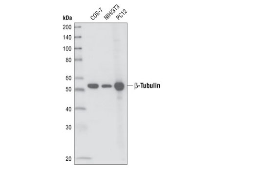
| β-Tubulin (9F3) Rabbit mAb 2128 | 20 µl |
|
H M R Mk Z B | 55 | Rabbit IgG |
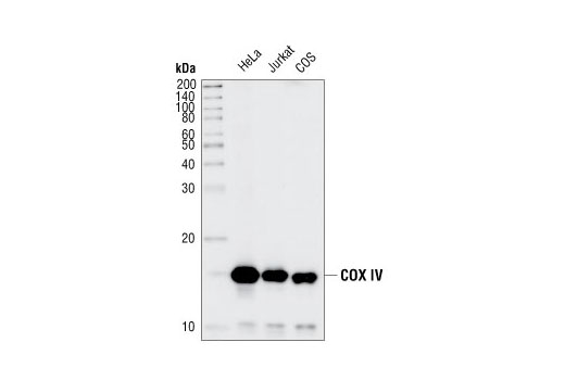
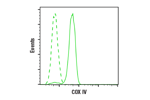
| COX IV (3E11) Rabbit mAb 4850 | 20 µl |
|
H R Mk Z B Pg | 17 | Rabbit IgG |
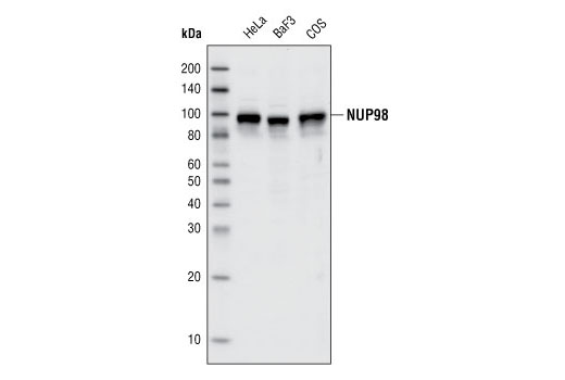
| NUP98 (C39A3) Rabbit mAb 2598 | 20 µl |
|
H M R Mk | 98 | Rabbit IgG |
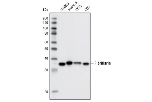
| Fibrillarin (C13C3) Rabbit mAb 2639 | 20 µl |
|
H M R Mk | 37 | Rabbit IgG |
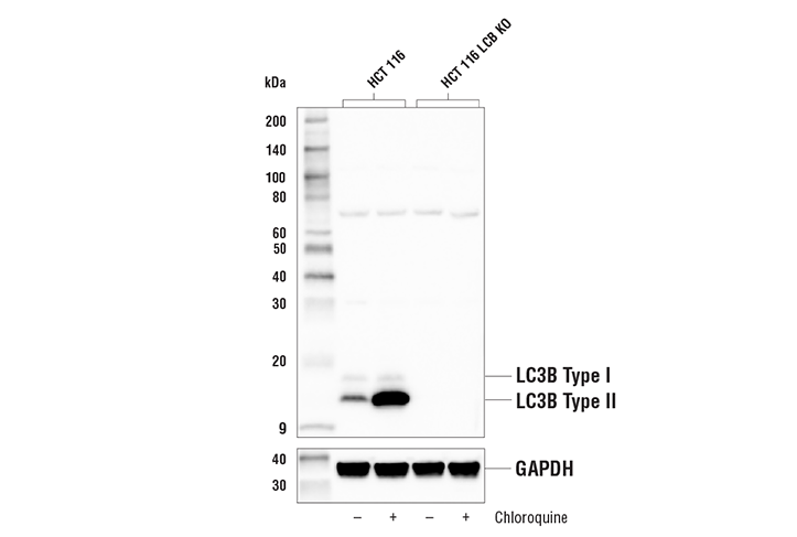
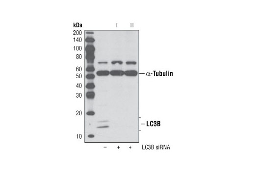
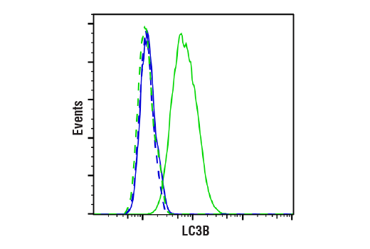
| LC3B (D11) XP® Rabbit mAb 3868 | 20 µl |
|
H | 14, 16 | Rabbit IgG |
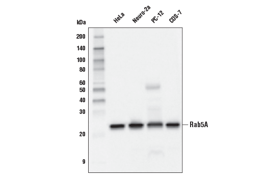
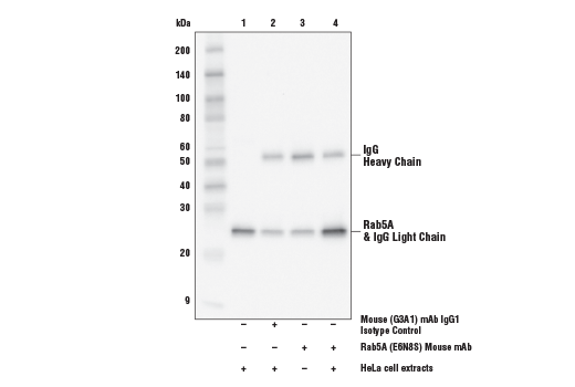
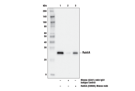
| Rab5A (E6N8S) Mouse mAb 46449 | 20 µl |
|
H M R Mk | 25 | Mouse IgG1 |
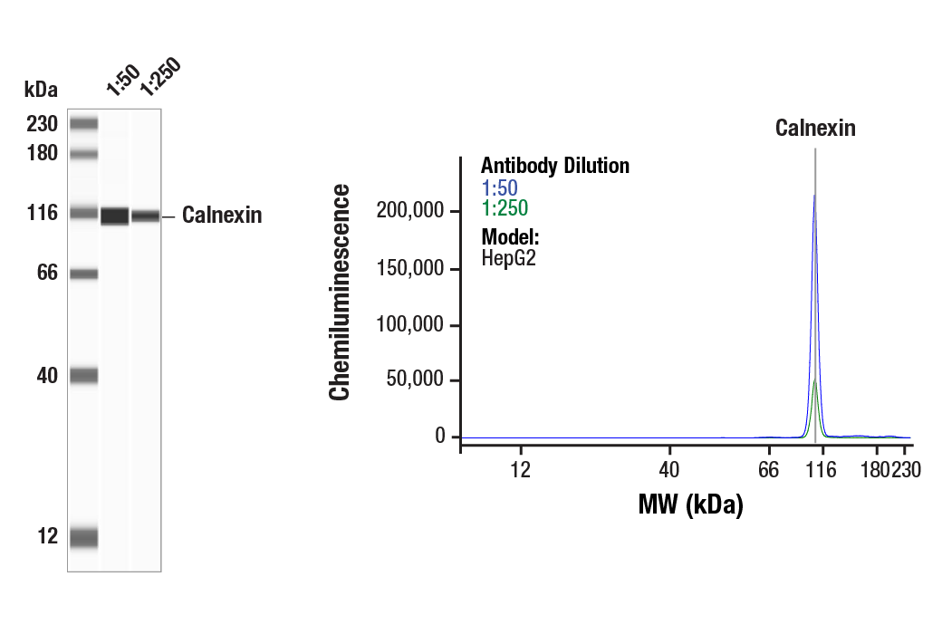
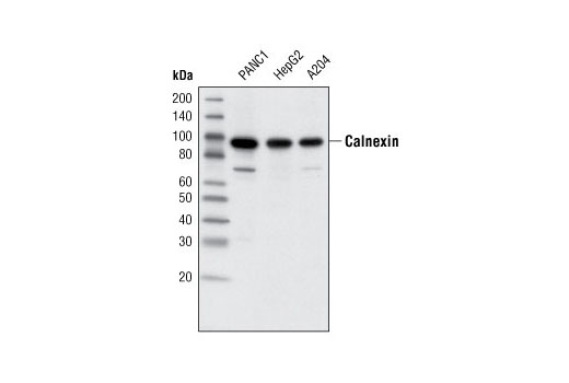
| Calnexin (C5C9) Rabbit mAb 2679 | 20 µl |
|
H Mk | 90 | Rabbit IgG |
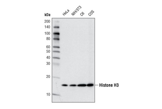
| Histone H3 (D1H2) XP® Rabbit mAb 4499 | 20 µl |
|
H M R Mk | 17 | Rabbit IgG |
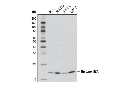
| Histone H2A (D6O3A) Rabbit mAb 12349 | 20 µl |
|
H M R Mk Z GP | 14 | Rabbit IgG |
Product Information
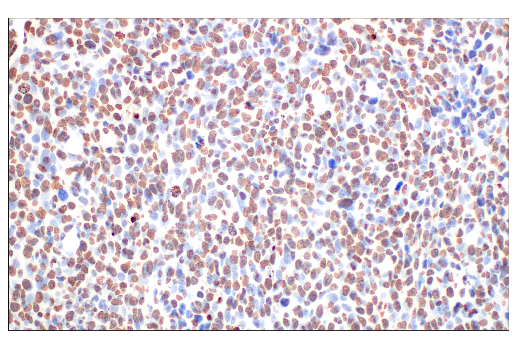
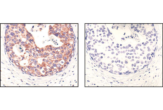
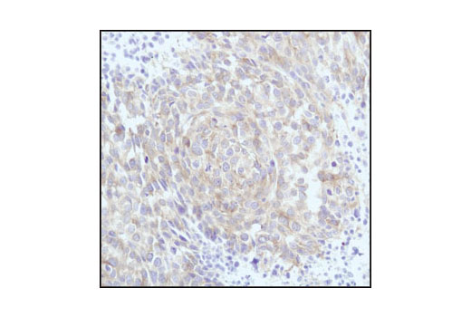
Rabbit monoclonal antibodies are prepared by immunizing animals with a synthetic peptide corresponding to: the amino terminus of human β-tubulin, the sequence of human calnexin, residues surrounding Lys29 of human COX IV, the carboxy-terminal sequence of human histone H3 and human histone H2A, residues surrounding Pro671 of human NUP98, residues surrounding Thr298 of human fibrillarin, and residues near the amino terminus of LC3B. Mouse monoclonal antibody is produced by immunizing animals with a synthetic peptide corresponding to residues surrounding Gly190 of human Rab5A protein.
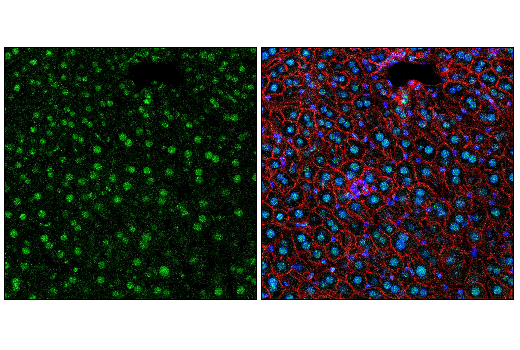
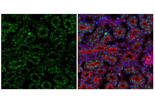
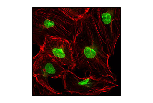
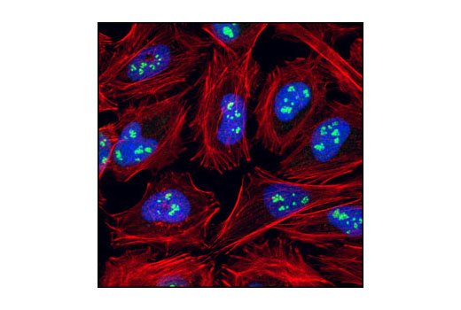
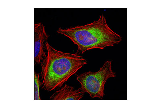
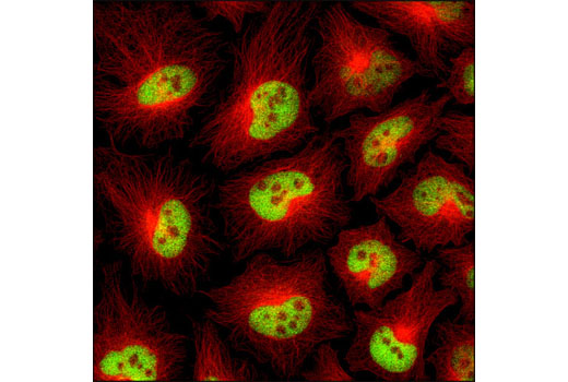
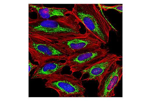
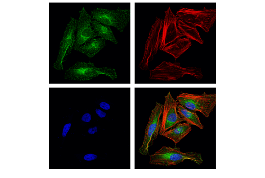
Knowledge of the subcellular location of a protein may reveal the potential role it plays in a variety of cellular processes. One can confirm the subcellular location of a marker that colocalizes with one of the organelle-specific antibodies in this kit. While these antibodies serve as powerful tools for immunofluorescence, they may also be used as western blot controls for fractionated cell lysates.
Except as otherwise expressly agreed in a writing signed by a legally authorized representative of CST, the following terms apply to Products provided by CST, its affiliates or its distributors. Any Customer's terms and conditions that are in addition to, or different from, those contained herein, unless separately accepted in writing by a legally authorized representative of CST, are rejected and are of no force or effect.
Products are labeled with For Research Use Only or a similar labeling statement and have not been approved, cleared, or licensed by the FDA or other regulatory foreign or domestic entity, for any purpose. Customer shall not use any Product for any diagnostic or therapeutic purpose, or otherwise in any manner that conflicts with its labeling statement. Products sold or licensed by CST are provided for Customer as the end-user and solely for research and development uses. Any use of Product for diagnostic, prophylactic or therapeutic purposes, or any purchase of Product for resale (alone or as a component) or other commercial purpose, requires a separate license from CST. Customer shall (a) not sell, license, loan, donate or otherwise transfer or make available any Product to any third party, whether alone or in combination with other materials, or use the Products to manufacture any commercial products, (b) not copy, modify, reverse engineer, decompile, disassemble or otherwise attempt to discover the underlying structure or technology of the Products, or use the Products for the purpose of developing any products or services that would compete with CST products or services, (c) not alter or remove from the Products any trademarks, trade names, logos, patent or copyright notices or markings, (d) use the Products solely in accordance with CST Product Terms of Sale and any applicable documentation, and (e) comply with any license, terms of service or similar agreement with respect to any third party products or services used by Customer in connection with the Products.
View in English?