View in English?
View in English?
View in English?

Product Information
Human
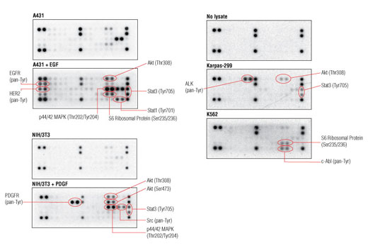
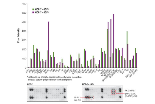
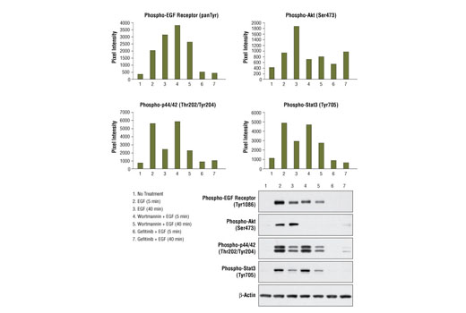
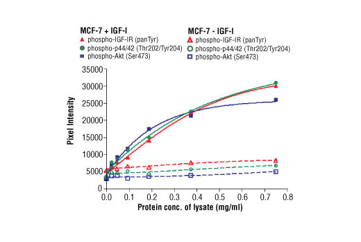
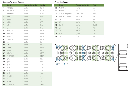
Receptor Tyrosine Kinases (RTKs) are a family of cell surface receptors that signal primarily through tyrosine phosphorylation events (1). RTKs trigger a wide range of downstream signaling cascades, including the PI3K/Akt, MAPK and Jak/Stat pathways. These pathways control basic cellular functions such as division, growth, metabolism, differentiation, migration and survival. Dysregulation of RTK signaling has been implicated in a large number of cancers (2), making RTKs popular targets for pharmaceutical intervention.
Explore pathways related to this product.
STRING - Known and Predicted Protein-Protein Interactions.
UniProt ID: P21709 , P40763 , P27361 , P29317 , P16234 , P35568 , P54753 , Q06418 , P04629 , P21860 , P31751 , Q9Y243 , P43403 , P07947 , P07948 , Q02763 , P30530 , P07333 , Q9UM73 , P62753 , P22455 , P28482 , P06239 , P00533 , P29320 , P31749 , P08581 , P11362 , P04626 , P10721 , P06213 , P54760 , P08631 , P07949 , P42224 , P08069 , Q16620 , P12931 , Q04912 , P35968 , P06241 , P00519
Entrez-Gene Id: 2041 , 6774 , 5595 , 1969 , 5156 , 3667 , 2049 , 7301 , 4914 , 2065 , 208 , 10000 , 7535 , 7525 , 4067 , 7010 , 558 , 1436 , 238 , 6194 , 2264 , 5594 , 3932 , 1956 , 2042 , 207 , 4233 , 2260 , 2064 , 3815 , 3643 , 2050 , 3055 , 5979 , 6772 , 3480 , 4915 , 6714 , 4486 , 3791 , 2534 , 25
Except as otherwise expressly agreed in a writing signed by a legally authorized representative of CST, the following terms apply to Products provided by CST, its affiliates or its distributors. Any Customer's terms and conditions that are in addition to, or different from, those contained herein, unless separately accepted in writing by a legally authorized representative of CST, are rejected and are of no force or effect.
Products are labeled with For Research Use Only or a similar labeling statement and have not been approved, cleared, or licensed by the FDA or other regulatory foreign or domestic entity, for any purpose. Customer shall not use any Product for any diagnostic or therapeutic purpose, or otherwise in any manner that conflicts with its labeling statement. Products sold or licensed by CST are provided for Customer as the end-user and solely for research and development uses. Any use of Product for diagnostic, prophylactic or therapeutic purposes, or any purchase of Product for resale (alone or as a component) or other commercial purpose, requires a separate license from CST. Customer shall (a) not sell, license, loan, donate or otherwise transfer or make available any Product to any third party, whether alone or in combination with other materials, or use the Products to manufacture any commercial products, (b) not copy, modify, reverse engineer, decompile, disassemble or otherwise attempt to discover the underlying structure or technology of the Products, or use the Products for the purpose of developing any products or services that would compete with CST products or services, (c) not alter or remove from the Products any trademarks, trade names, logos, patent or copyright notices or markings, (d) use the Products solely in accordance with CST Product Terms of Sale and any applicable documentation, and (e) comply with any license, terms of service or similar agreement with respect to any third party products or services used by Customer in connection with the Products.
View in English?