View in English?
View in English?
View in English?

| Cat. # | Size | Qty. | Price | Inventory |
|---|---|---|---|---|
| 9956T | 1 Kit (7 x 20 microliters) |
|
| Product Includes | Quantity | Applications | Reactivity | MW(kDa) | Isotype |
|---|---|---|---|---|---|
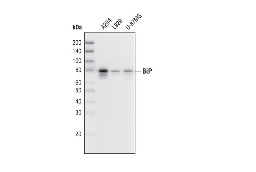
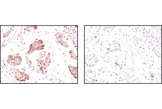
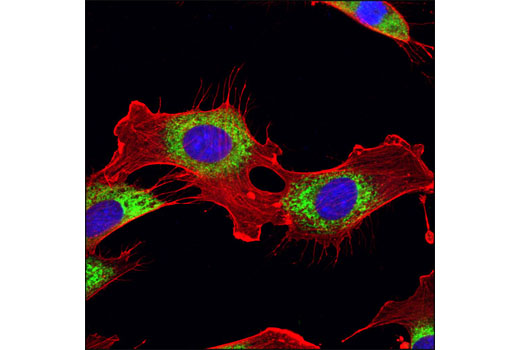
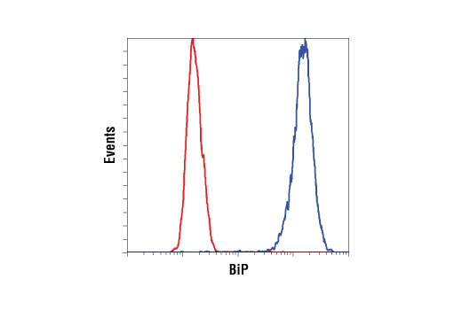
| BiP (C50B12) Rabbit mAb 3177 | 20 µl |
|
H M | 78 | Rabbit IgG |
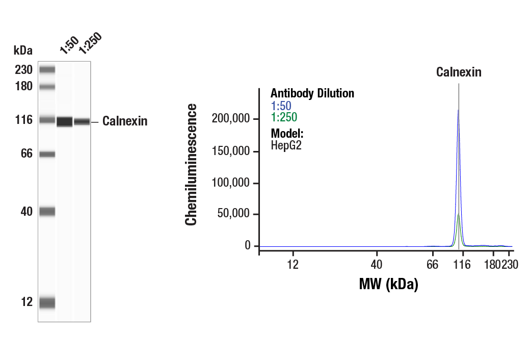
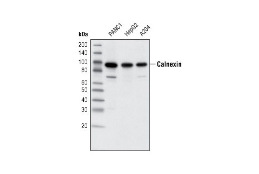
| Calnexin (C5C9) Rabbit mAb 2679 | 20 µl |
|
H Mk | 90 | Rabbit IgG |
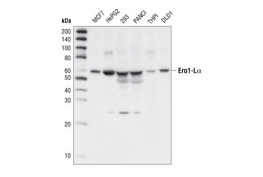
| Ero1-Lα Antibody 3264 | 20 µl |
|
H | 60 | Rabbit |
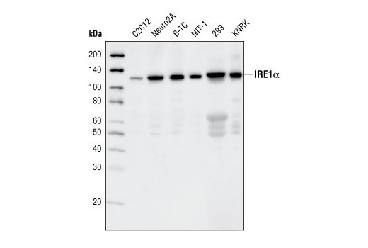
| IRE1α (14C10) Rabbit mAb 3294 | 20 µl |
|
H M R | 130 | Rabbit IgG |
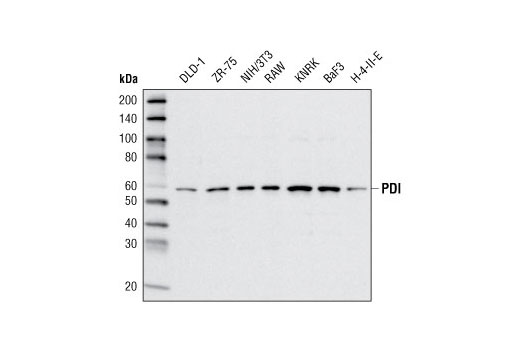
| PDI (C81H6) Rabbit mAb 3501 | 20 µl |
|
H M R Mk | 57 | Rabbit |
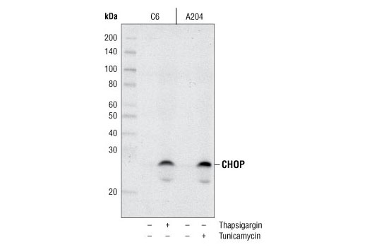
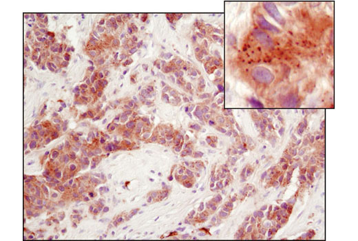
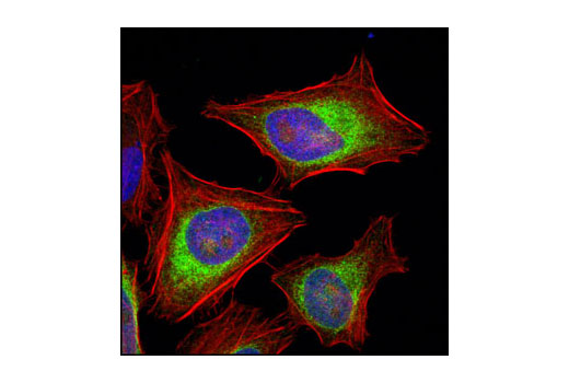
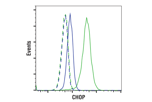
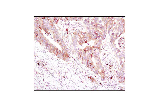
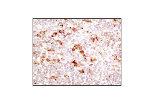
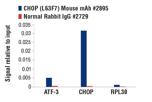
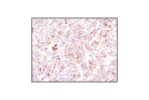
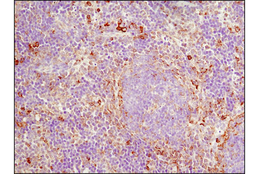
| CHOP (L63F7) Mouse mAb 2895 | 20 µl |
|
H M R | 27 | Mouse IgG2a |
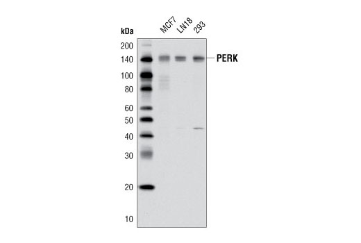
| PERK (D11A8) Rabbit mAb 5683 | 20 µl |
|
H | 140 | Rabbit IgG |
| Anti-rabbit IgG, HRP-linked Antibody 7074 | 100 µl |
|
Goat | ||
| Anti-mouse IgG, HRP-linked Antibody 7076 | 100 µl |
|
Horse |
Product Information
Monoclonal antibody is produced by immunizing animals with a synthetic peptide corresponding to residues surrounding Leu156 of human PERK protein, the sequence around Gly584 of human BiP, the sequence around His963 of human IRE1α, the sequence of human PDI and the sequence of human CHOP. Polyclonal antibodies are produced by immunizing animals with a synthetic peptide corresponding to a sequence around Ala51 of human calnexin, the sequence around Leu218 of human Ero1-Lα, and the sequence of mouse MBTPS2. Antibodies are purified by protein A and peptide affinity chromatography.
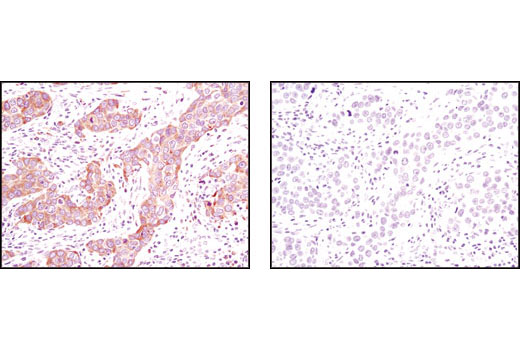
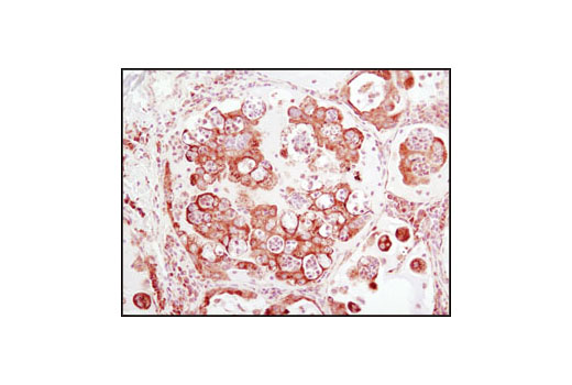
Secretory and transmembrane proteins are synthesized on polysomes and translocate into the endoplasmic reticulum (ER) where they are often modified by the formation of disulfide bonds, amino-linked glycosylation and folding. The ER contains a pool of molecular chaperone proteins including calnexin, BiP and protein disulfide isomerase (PDI). Calnexin is an ER membrane, calcium-binding protein that retains newly synthesized glycoproteins inside the ER to ensure proper folding and quality control (1,2). Irregular protein folding within the ER increases BiP synthesis, which binds misfolded proteins to prevent them from forming aggregates and to assist them to refold properly (3).
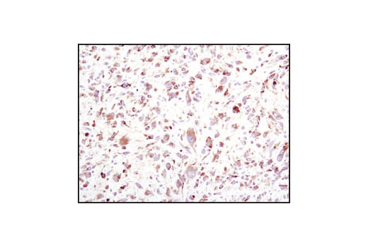
PDI catalyzes the formation and isomerization of disulfide bonds required for a protein to reach its native state (4). Studies have found that the resident ER protein endoplasmic oxidoreductin-1 (Ero1) provides oxidizing potential to the ER in Saccharomyces cerevisiae (5). Ero1-Lα is an ER membrane-associated N-glycoprotein that promotes oxidative protein folding (6). Disruptions of ER homeostasis leads to the accumulation of unfolded proteins. The ER has developed an adaptive mechanism called the unfolded protein response (UPR) to counteract compromised protein folding (7). This is regulated by proteins such as the membrane-bound transcription factor protease site 2 (MBTPS2) and the serine/threonine kinase IRE1 (8-12). The PERK eIF2α kinase is an ER resident transmembrane protein that couples ER stress signals to translation inhibition. ER stress increases PERK activity, which phosphorylates eIF2α to reduce protein translation. PERK activation during ER stress correlates with autophosphorylation of its cytoplasmic kinase domain (13,14). Phosphorylation of PERK at Thr980 can serve as a marker for its activation status.
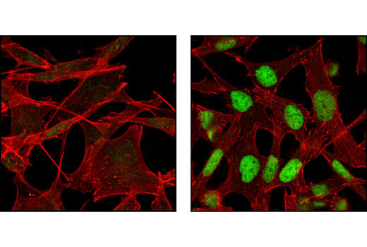
During ER stress, the level of CHOP expression is elevated and CHOP functions to mediate programmed cell death (15).
Explore pathways related to this product.
STRING - Known and Predicted Protein-Protein Interactions.
Except as otherwise expressly agreed in a writing signed by a legally authorized representative of CST, the following terms apply to Products provided by CST, its affiliates or its distributors. Any Customer's terms and conditions that are in addition to, or different from, those contained herein, unless separately accepted in writing by a legally authorized representative of CST, are rejected and are of no force or effect.
Products are labeled with For Research Use Only or a similar labeling statement and have not been approved, cleared, or licensed by the FDA or other regulatory foreign or domestic entity, for any purpose. Customer shall not use any Product for any diagnostic or therapeutic purpose, or otherwise in any manner that conflicts with its labeling statement. Products sold or licensed by CST are provided for Customer as the end-user and solely for research and development uses. Any use of Product for diagnostic, prophylactic or therapeutic purposes, or any purchase of Product for resale (alone or as a component) or other commercial purpose, requires a separate license from CST. Customer shall (a) not sell, license, loan, donate or otherwise transfer or make available any Product to any third party, whether alone or in combination with other materials, or use the Products to manufacture any commercial products, (b) not copy, modify, reverse engineer, decompile, disassemble or otherwise attempt to discover the underlying structure or technology of the Products, or use the Products for the purpose of developing any products or services that would compete with CST products or services, (c) not alter or remove from the Products any trademarks, trade names, logos, patent or copyright notices or markings, (d) use the Products solely in accordance with CST Product Terms of Sale and any applicable documentation, and (e) comply with any license, terms of service or similar agreement with respect to any third party products or services used by Customer in connection with the Products.
View in English?